The Renal Pedicle: A Surgeon's Anatomical Guide
Wiki Article

The kidneys are vital organs, and their successful surgical management hinges on a deep understanding of their complex anatomy. At the heart of this anatomy is the renal pedicle, a critical bundle of structures that serves as the kidney's lifeline. For surgeons, medical students, and healthcare professionals, mastering the intricacies of this area is not just an academic exercise—renal pedicle is essential for procedural success and patient safety. A clear grasp of its components and variations is fundamental to performing procedures ranging from nephrectomies to renal transplants with precision and confidence.
Understanding the Core Components
The renal pedicle refers to the collection of vessels and structures that enter and exit the kidney at its hilum. Primarily, this includes the renal artery, the renal vein, and the renal pelvis, which funnels urine from the kidney to the ureter. Lymphatic vessels and autonomic nerves also travel within this bundle. The precise arrangement of these structures can vary, but typically, the renal vein is the most anterior structure, followed by the renal artery, with the renal pelvis positioned most posteriorly. This anatomical relationship is a crucial landmark for surgeons navigating this delicate region.
Surgical Significance and Procedural Nuances
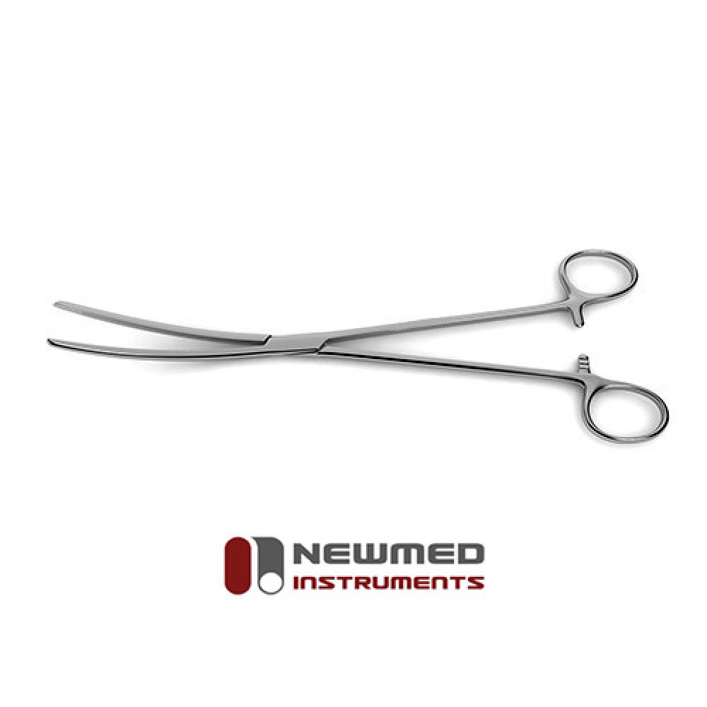
Nearly every major kidney operation involves interaction with the renal pedicle. During a nephrectomy, for instance, the surgeon must carefully dissect and ligate the renal artery and vein to remove the kidney safely with micro clamps while preventing catastrophic hemorrhage. In kidney transplantation, controlling and preparing the renal pedicle of the donor organ is a key step for ensuring a successful anastomosis in the recipient. The complexity of these procedures demands not only skill but also instruments that provide flawless performance. At New Med Instruments, we are committed to providing surgeons with the quality tools they need to navigate these challenges, reflecting our dedication to superior service and enabling perfect, precise results for every patient.
Navigating Anatomical Variations
While textbooks provide a standard model, surgeons must be prepared for anatomical variations of the renal pedicle. The presence of accessory renal arteries, which occur in about 30% of individuals, is a common example. These additional arteries can arise from the aorta or other nearby vessels and must be identified and managed to avoid complications like segmental ischemia or bleeding. Variations in the renal vein, such as multiple veins or retroaortic positioning, also present unique challenges. Acknowledging and preparing for these potential differences are hallmarks of an experienced surgeon.
The Role of High-Quality Instrumentation
Precision is paramount when working around such a critical structure. The instruments used must offer exceptional control, reliability, and tactile feedback. Whether a surgeon is meticulously dissecting delicate tissues, ligating major vessels, or performing a complex reconstruction, the quality of their tools directly impacts the outcome. New Med Instruments was founded on the principle of equipping healthcare professionals with superior surgical instruments. We support surgeons, whether they are just starting their practice or are seasoned experts, by providing the precise tools needed to manage the renal pedicle and perform even the most demanding procedures with confidence.
Report this wiki page